Dental & Vision
Why Diabetes Shows Up In The Retina First
The back of the eye is the only place where small blood vessels can be seen directly, which makes routine eye examination an early window on damage happening elsewhere.
People with diabetes are told to have their eyes examined regularly even when their vision seems fine. The reason is anatomical as much as ophthalmic.
Small vessels are damaged before large ones
Persistently elevated blood glucose damages the walls of the smallest blood vessels, weakening their structure and making them leak and close off over time.
Tissues that depend on dense networks of these vessels, including the retina, the kidney's filtering units and the nerves in the extremities, are affected earliest.
Damage in all three sites reflects the same underlying process, which is why a finding in one raises the likelihood of changes in the others.
The retina is uniquely visible
Nowhere else in the body can a clinician look directly at living capillaries without an incision, because the eye's optics provide a clear path to the retinal surface.
Through a dilated pupil or a retinal camera, individual vessels, small hemorrhages and leaked material can be seen and photographed in a few minutes.
That accessibility turns an eye examination into a low-cost observation of microvascular health rather than a test only about sight.
Early changes do not affect vision
The first visible signs are small outpouchings in vessel walls and tiny areas of leakage, located away from the central region responsible for detailed vision.
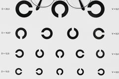
A person can therefore have clearly documented retinal changes while reading a standard eye chart perfectly, which is the central reason screening is scheduled rather than symptom-driven.
By the time vision is noticeably affected, the process has usually reached the central retina or triggered the growth of fragile new vessels.
Screening is organized around that gap
Diabetic eye screening programs work on defined intervals, with images graded and people recalled sooner when specific features appear rather than when symptoms are reported.
Retinal photography allows the images to be captured in one setting and assessed elsewhere, which is what makes population-scale screening practical.
Recommended intervals depend on the type of diabetes, how long it has been present and what previous images showed, and they are set by the treating team.
The finding feeds back into general care
Retinal changes are used as evidence about how the whole vascular system is faring, and they inform decisions about overall management rather than eye treatment alone.
Blood pressure, blood glucose control and kidney function are all part of that picture, and eye findings are shared with the clinicians handling them.
What any individual result means, and what should follow it, is a matter for the ophthalmologist and the diabetes team who hold the rest of the record.
Also by Dr Samuel Adeyemi
- Dental emergencies and what to doDental & Vision
- Overdiagnosis, and why more medicine is not always betterScreening & Checks
- Mouth ulcers, bad breath and the small thingsDental & Vision
- Dry eye, contact lenses and daily eye careDental & Vision