Dental & Vision
What A Dental X-Ray Shows That Looking Cannot
Radiographs reveal decay between teeth, bone levels and structures below the gum, all of which are invisible to direct examination however careful it is.
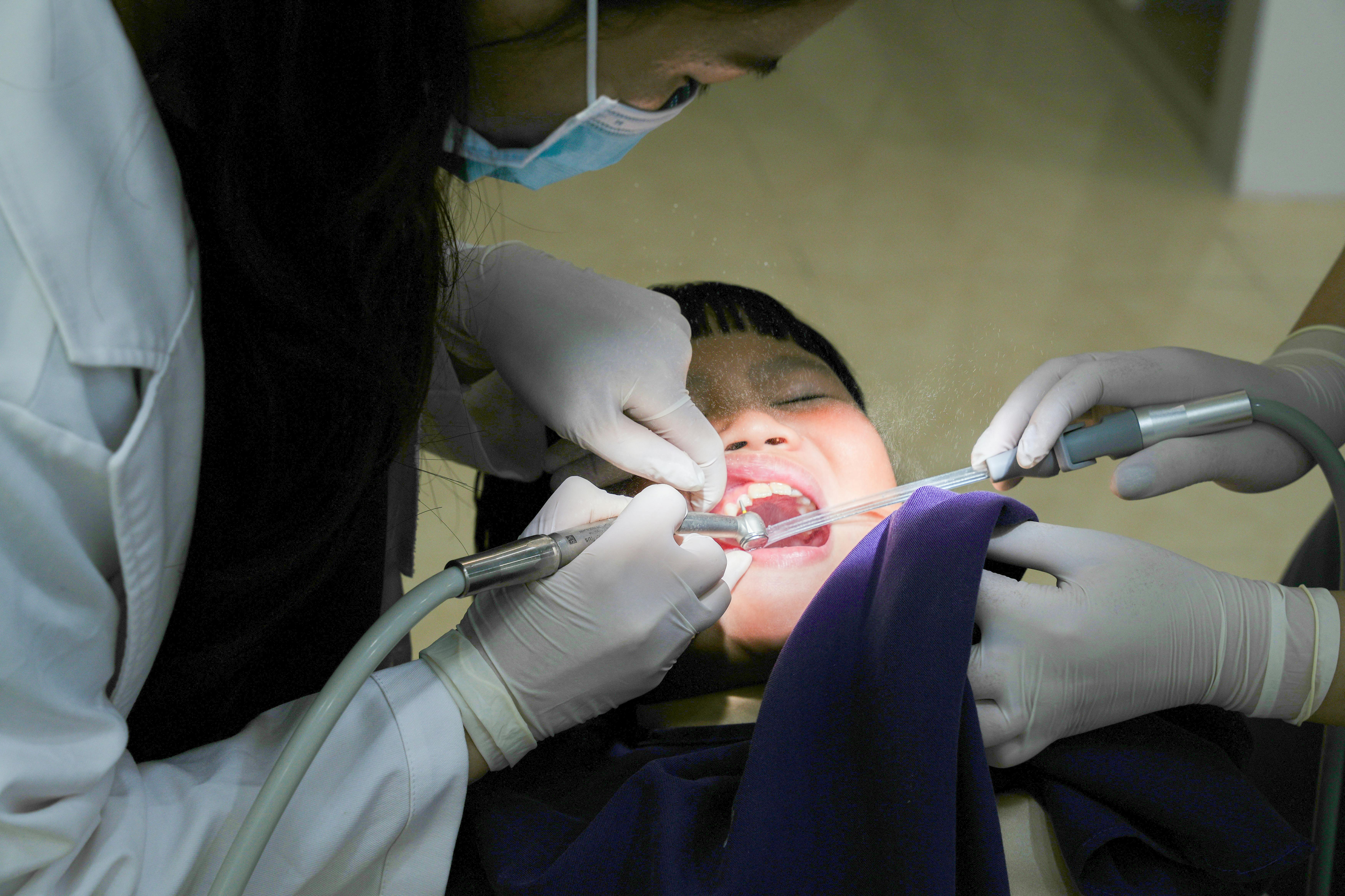
Dental radiographs are often assumed to be a routine formality. They image regions that no amount of looking can reach, and they are the reason some problems are found before they are felt.
Contact surfaces are hidden by geometry
Adjacent teeth touch each other, and the surfaces at that contact point cannot be seen or probed while both teeth are present.
Decay commonly starts exactly there, because the contact area traps plaque and resists brushing, and floss reaches it only partially.
An X-ray passes through the teeth and reveals the density change caused by mineral loss, which is why interproximal decay is almost always a radiographic finding.
Bone level is the key periodontal measurement
Radiographs show the height of bone around each root, which records how much support has been lost over time.
Probing depths measure the soft tissue picture, and the two together give a clearer account than either alone.
Comparing images taken years apart shows the direction of travel, which matters far more clinically than any single measurement in isolation.
Roots and developing teeth are invisible otherwise
Infection at the tip of a root produces a change in the surrounding bone that appears on an image long before it causes swelling or pain.
In children, radiographs show whether adult teeth are present, correctly positioned and developing on schedule, which informs decisions about extraction and orthodontics.
Impacted teeth, cysts and retained roots are found the same way, generally as incidental findings during examination for something else.
Restorations are checked from underneath
Decay can develop beneath an existing filling or crown, where the restoration itself blocks any view of the tooth structure below.
Radiographs also show whether a restoration margin is sealed and whether a root filling extends properly along the canal.
This is why images are taken before certain treatments rather than only when a problem is suspected, since the plan depends on what lies underneath.
Dose and frequency are governed deliberately
Dental radiographs use low doses, and digital sensors have reduced them further compared with film.
Frequency is set by individual risk rather than by a fixed schedule, so someone with a history of decay is imaged more often than someone with none.
Anyone unsure why an image is being taken can ask what question it is expected to answer, which is a reasonable request and one the dentist should be able to state plainly.
Also by Renata Fiore
- Deciding what to spend on your healthBills & Access
- Workplace health benefits people never useBills & Access
- Choosing between public and private treatmentBills & Access
- End-of-life planning and its practical costsBills & Access